Questions on Analytical chemistry (SL & HL)

A.1 Analytical techniques - questions

1 - What are the different types of analysis ?

2 - What methods can be used to give information about the molecule structure ?

3 - Summarize all the methods !

A.2 Spectroscopy - questions

3 - Describe what electromagnetic radiation is.

4 - What is the formula for the energy and wavelength of a photon ?

5 - What is the wavenumber ?

6 - Describe the electromagnetic spectrum.

7 - What is an absorption and emission spectrum ?

A.3 IR Spectroscopy - questions

8 - How can infrared radiation be used to excite a molecule ?

9 - What sort of movement in the molecule can the IR radiation induce ?

10 - How does the wavenumber depend on the bond type in diatomic molecules ?

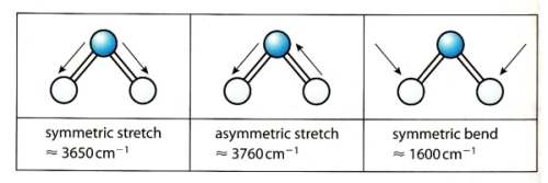
11 - How does the wavenumber depend on the vibrational mode in polyatomic molecules ?

12 - Only two vibrational modes are active for the carbon dioxide molecule. Why ?

13 - What are the characteristic wavenumber for different bonds ?

14 - Explain how a double-beam IR spectrometer works !

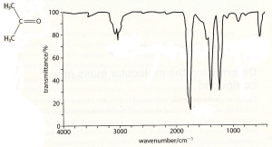
15 - Analyse  

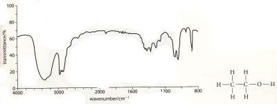
16 - Analyse  
A.4 Mass Spectroscopy - questions

17 - Describe how a mass spectrometer works.

18 - Describe how a mass spectrometer can be used to get information about the molecular spectrum.

19 - Solve  

A.5 & A.9 NMR Spectroscopy - questions

20 - Describe how a NMR spectrometer works.

21 - What is the chemical shift ?

22 - What are the characteristic chemical shifts for hydrogen atoms in different chemical environments ?

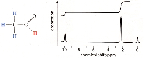
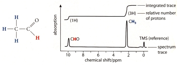
23 - What is shown here:  
24 - NMR spectrum of C3H8O:  
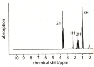
Which formula is correct:  

25 - What is MRI ?

26 - Why is TMS used as a reference sample ?

27 - What would this look like in high-resolution NMR:
  

28 - What is causing the splitting of the peaks ?

29 - Explain the details of the NMR spectrum above.

30 - What is the different height of the peaks caused by ?

A.6 AA Spectroscopy - questions

31 - How does AA spectroscopy work ?

32 - What is a calibration curve and how it is used ?

A.8 UV Spectroscopy - questions

33 - What is a ligand ?

34 - How does ligands affect the energy levels of transition metals ?

35 - Why does transition metals with ligands such as [Cu(H2O)6]2+ appear coloured ?

36 - How does UV spectroscopy work ?

37 - What factors affect the colour of transition metal complexes ?

38 - What is a conjugated system ?

39 - What is a chromophore ?

40 - Why are carrots not blue ?


41 - Why does the colour in acid-base indicators change ?

42 - Explain how one can use UV-vis spectroscopy and Beer-Lamberts law to measure the concentration of metal ions !

A.7 & A.10 Chromatography - questions

43 - What is chromatography ?

44 - What is absorption and adsorption ?

45 - Explain the difference between adsorption chromatography and partition chromatography !

46 - Give examples of adsorption chromatography and partition chromatography !

47 - Explain how paper chromatography works !

48 - What is the retention factor ?

49 - Explain how Thin-Layer Chromatography (TLC) works !

50 - What are the advantages of TLC compared to paper chromatography ?

51 - Explain how column chromatography works !

52 - What are the advantages of column chromatography ?

53 - Explain how gas-liquid chromatography (GLC) works !

54 - Explain how the detector in GLC works !

55 - What is the difference in the chromatogram in GLC compared to that of TLC and paper chromatography ?

56 - When is gas-liquid chromatography used ?

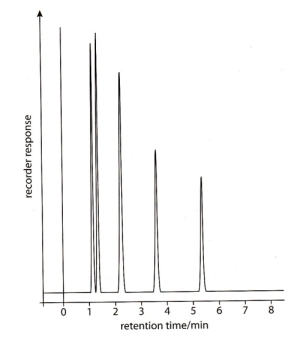
57 - What alcohols are causing these GLC peaks:
  

58 - Explain how High-Performance Liquid Chromatography (HPLC) works !

59 - What are the advantages of HPLC ?


Go back to the IB chemistry page

Go to the IB physics page
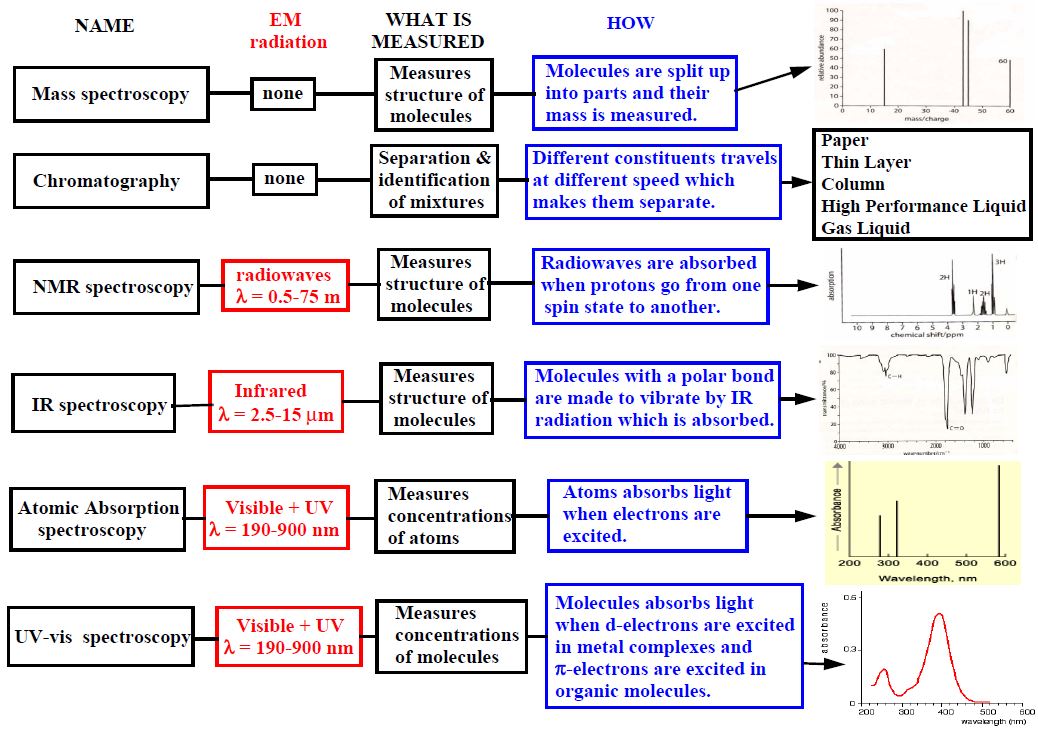
A.1 Analytical techniques


Qualitative analysis: The detection of the presence but not quantity of a chemical.
Quantitative analysis: The measurement of the quantity of a substance in a mixture.
Structural analysis: Measurement of how the atoms are arranged in a structure in the molecules.


IR Spectroscopy: Can be used to identify bonds in a chemical.
Mass Spectroscopy: Can be used to determine relative atomic and molecular masses.
NMR Spectroscopy: Can show the chemical environment for certain isotopes in a molecule.


A.2 Spectroscopy


Electromagnetic radiation is form of energy transferred by waves that are described by:
Wavelength (λ): The distance between to peaks.
Frequency (f): The number of waves passing a point every second.
The energy of electromagnetic radiation is carried in packets called photons.


E = h • f (LOOK IN THE DATA BOOKLET !)

Where h is the Planck's constant = 6.63• 10-34 Js (LOOK IN THE DATA BOOKLET !)

λ = c/f

Where c is the speed of light = 3 • 108 m/s

So the energy of electromagnetic radiation depends only on its frequency (or wavelength). It does not depend on the intensity of the radiation. This means for example that strong sun light photons do not have more energy than photons from weak moon light.


The wavenumber is the inverse of the wavelength. Its unit is cm-1

Wavenumber = 1 / λ


Radio waves: Can be absorbed by certain nuclei causing them to reverse their spin.
This is used in NMR and can give information about the environment of certain atoms.

Micro waves: Cause molecules to increase their rotational energy. Can give information about bond length.

Infrared waves: Is absorbed by certain bonds causing them to stretch or bend.

Visible and ultraviolet light: Can produce electronic transitions. Can give information about energy levels.

X rays: The wavelength is similar to the distance between atoms in crystals.
Can produce diffraction patterns which give information about atomic structure.

Gamma rays: Can cause changes in the energy of atomic nuclei. Not used in analytical chemistry.


Emission spectrum: If atoms in a gas is excited the electrons in the gas are going to higher energy levels. When they fall back to the groundstate they can send out the energy difference between the levels as light. Every energy difference has a specific energy or wavelength. The spectrum of this light has lines that corresponds to the levels.

Absorption spectrum: If instead white light with all different wavelengths is sent though a gas the atoms in the gas can get excited. Only light with wavelengths that corresponds exactly to the energy levels is absorbed. The emission spectrum has therefore black lines.


A.3 IR Spectroscopy


If a bond in a molecule is polar, the molecule has parts which are more positively charged and more negatively charged. In this case an electromagnetic wave with exactly the right IR frequency (= the natural frequency of a bond) can excite the bond between the atoms so that the molecule start to vibrate. The electromagnetic wave is then absorbed by the molecule.


Diatomic molecules: Can only vibrate by stretching the bond between the atoms.

Polyatomic molecules: The bonds can also bend.


Light atoms vibrate at higher frequency (have larger wavenumber) than heavy atoms.

Wavenumber of single bonds > Wavenumber of triple bonds > Wavenumber of double bonds


Wavenumber of asymmetric stretch > Wavenumber of symmetric stretch > Wavenumber of symmetric bend


LOOK IN THE DATA BOOKLET !


1. IR radiation from a heated filament is split into two parallel beams.

2. The radiation has some characteristic frequencies absorbed by a sample and a reference sample.

3. The beams through the sample and the reference are measured by a detector.

4. The differences in the intensities of the two beams is recorded as a function of the wavenumber.

5. When the radiation is absorbed at characteristic wavenumbers the transmission goes down and the curve goes down.

6. The purpose of the reference is to eliminate dips in the curve due to CO2 and H2O in the air or absorptions by the solvent used.

7. Molecules with several bonds can vibrate in many ways and give a complex pattern that can be used as a fingerprint and compared to known patterns.


LOOK IN THE DATA BOOKLET !


LOOK IN THE DATA BOOKLET !


A.4 Mass Spectroscopy


The stages of operation are: vaporisation, ionisation, acceleration, deflection and detection.

Substance to be tested is vaporised (by heat, in the absence of oxygen) then ionised by electric current. Ions are accelerated through an electric field, then deflected by a magnetic field. Ions are then detected. The angle of deflection due to the magnetic field reflects their mass to charge ratio.

The angle of deflection of each fragment is proportional to its mass, and so it is possible to find the relative atomic mass of each 'spike'. The height of the spike represents the frequency, therefore, the abundance can be calculated. The relative atomic mass is the weighted average of the isotope masses times their percentage abundance (frequencies).


The principle is that when the electrons hit the molecule in the mass spectrometer it will sometimes only ionize the molecule (which is then called a parent ion) but sometime it will break the molecule up into fragments. Different fragmentation ions give rise to a fragmentation pattern that depends on their relative mass and this pattern can give information about the molecules structure.


A.5 NMR Spectroscopy


1. Atoms with odd number of protons can be in two spin states with different energy.

2. Examples of such atoms is the most common hydrogen isotope 1H and the rare carbon isotope 13C but not 12C.

3. Normally half the atoms are randomly in one or the other of the two spin states but if they are put in a magnetic field they all end up in the state with the lowest energy.

4. If a radio frequent electromagnetic field with exactly the right resonance frequency is applied then all the atoms can go from one spin state to the other.

5. When this happens a signal is created which is picked up and analyzed by the nuclear magnetic resonance spectrometer.


1. The electrons in a molecule will shield the nucleus from the effect of the external magnetic field.

2. This means that hydrogen atoms will have slightly different resonance signals depending on where in the molecule they are.

3. The signals from a sample is compared to that of tetramethylsilane (TMS) which is used as a reference.

4. The difference of the position of the NMR signal to that of TMS is called the chemical shift.


LOOK IN THE DATA BOOKLET !


A NMR Spectrum.

The top curve shows the integrated value. The increase at the first peak is 1/3 of the increase at the second peak. The fact that the integral of the peak at 2 is three times larger indicates that there are 3 times as many hydrogen atoms in that chemical environment than for the peak at 10. A look in the data booklet shows that CHO has a chemical shift of 9.7 so the first peak is that. CH3 bonded to a C with a bond to O has 2.1 so that is the second peak. The peak at 0 is always from TMS.


The formula to the left is correct because it has hydrogen atoms in four different chemical environments (I, II, III and IV) while the others have only 3 (I, II, III).


Location of the peaks can be identified from the chemical environments I, II, III and IV by looking in the Data Booklet:
I: R-CH3 at 0.9
II: R-CH2-R at 1.3
III: R-CH2-hal at 3.2-3.7
IV: R-O-H at 0.5-6.5


MRI stands for Magnetic Resonance Imaging. It as a medical diagnostic tool that uses NMR on the water in the human body. People are put in a strong magnetic field and bombarded by radiopulses. The radiowaves are then detected and computers are used to produce images.


A.9 Further NMR Spectroscopy


TMS or tetramethylsilicane is used as a reference because:

1. All hydrogen nuclei in TMS is in the same environment which gives only one peak.

2. Silicon has a lower electronegativity than Carbon and so TMS gives a peak in a different region from molecules with C-H bonds.

3. The frequency of the absorbed radiowaves in NMR depends on the strength of the magnetic field but not the chemical shift with respect to TMS.

4. TMS is chemically inert and is soluble in most organic solvents.

5. TMS has a low boiling point and can therefore be easily removed from a sample.


Compare a low-resolution NMR spectrum with a high-resolution one:


The peaks in a NMR spectrum depend not only on the external magnetic field but also on the magnetic field created by the closest Hydrogen neighbours. If a Hydrogen atom has one other close Hydrogen neighbour atom then the peak is split in two because the magnetic field of the neighbour can be in two directions. If it has two Hydrogen neighbour atoms the peak is split into three peaks. If it has three Hydrogen neighbour atoms the peak is split into four peaks. So the number of peaks is equal to the number of neighbours + 1.


The height of the peaks depends on the number of different combinations the spin of the neighbour hydrogen atoms can be in.
This is given by the Pascal triangle:


A.6 AA Spectroscopy


1. With atomic absorption spectrometry you want to know how much of a particular element there is in a sample.

2. A special lamp is made using the element one is looking for. This lamp is giving a particular emission spectrum that depends on the element.

3. The light from the special lamp is going through a flame made of the sample under study, some fuel and air.

4. If there is the same element in the lamp and the flame then the emission light will be absorbed in the flame.

5. A monochromator is used to select a particular wavelength to study and a photomultiplier is used to measure the intensity of the light.

6. A reference beam that did not go through the flame is also measured.

7. The ratio of the intensity of the light through the flame and not through the flame gives a measure of how much of the element is in the sample.


A calibration curve is made by doing measurements with samples with different known concentrations. A measurement with a sample with unknown concentration can then be estimated using the calibration curve. An example is given below for a sample with 25% absorption of light. The curve shows that this corresponds to a concentration of 3 micrograms per liter.


A.8 UV Spectroscopy


A ligand is a molecular group such as H2O, NH3, OH- or CO that has at least one atom with a lone pair of electrons which is used to form a covalent bond with a central metal ion. An example of a metal compound with 6 H2O ligands is [Cu(H2O)6]2+ .


The outer electrons of transition metals are in d-orbitals. There are 5 different d-orbitals and normally they have all the same energy levels.
Cu2+ has for example nine electrons in five d-orbitals that all have the same energy.
However, the 6 H2O ligands in [Cu(H2O)6]2+ will affect the energy levels of the d-orbitals so that two groups of orbitals are created with different energy. This means that the energy level of the two orbitals to the left in the figure below is higher than the energy level of the three orbitals on the right.


When light passes through a solution of [Cu(H2O)6]2+ then orange light is absorbed and used to excite one electron from a low to a high d-orbital energy level. When the orange wavelength in the light disappear the remaining light looks turquoise because orange is on the opposite side of turquoise in the color wheel:


UV and visible light is sent through a solution of complex metal ions or organic compounds. Some of the light is absorbed due to d-orbital excitation. The principle is the same as in IR-spectroscopy but in the measurement one calculates wavelengths instead of wavenumber and absorption rather than transmission. The absorbed spectrum does not have sharp peaks but broad dips since the ligands have rotational and vibrational energy which means that they can take up excess energy in the absorption process.


Nuclear charge of central metal ion: The strength of the bond between the metal ion and the ligand depends on this charge and this affects the difference in energy levels which in turn affects the color.

Charge density of the ligand: A ligand with a larger charge density cause a larger split in the energy levels and the wavelength were maximum absorbance occur decreases with increasing charge density.

The geometry of the complex ion: The energy levels (or the splitting of the d-orbitals) depends on the orientation of the ligands.

Number of electrons: The strength of the interaction between the ligand and the metal ion depends also on the number of d-electrons. This affects the splitting of energy levels and thus the color.


A conjugated system is when you have alternating single and double bonds C=C-C=C-C=C-C=C. This creates a system of connected p-orbitals with delocalized pi electrons.


A chromophore is the part of a molecule responsible for its color. The color arises when a molecule absorbs certain wavelengths of visible light and transmits or reflects others. The chromophore is a region in the molecule where the energy difference between two different molecular orbitals falls within the range of the visible spectrum. Visible light that hits the chromophore can thus be absorbed by exciting an electron from its ground state into an excited state.

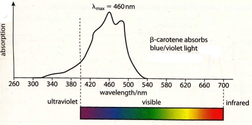
In the conjugated chromophores, the electrons jump between energy levels that are extended pi orbitals, created by a series of alternating single and double bonds, C=C-C=C. The longer the chain, the higher the wavelength of the absorbed light is.


The UV-vis spectrum of β-carotine shows that it absorbs blue/violet light. So one has to go to the opposite side of the colour wheel to see that its colour is orange:


This is because when you add H+ or OH- ions to the indicators the degree of conjugation changes in their molecules. This changes the energy levels and thus the wavelength at which light is absorbed.


1. Light with a wavelength that is known to be absorbed by the metal ions in the sample is selected by the monochromator.

2. The intensity (Io) of this light is measured without a sample.

3. A sample is installed in the spectrometer and the intensity (I) of the light going through the sample is measured.

4. Lambert-Beer's law then says that A = log(I/Io) = ε c L

5. L is the path length or the length of the container with the sample. This length is known.

6. A is the absorbance. This is what is being measured.

7. ε is the molar absorptivity which is the absorbance of a 1 mol/dm3 solution in a 1 cm container.

9. c is the concentration that one is trying to measure.

10. In practice one is not using the Lambert-Beer's law as a formula. Instead one puts several solutions with known concentration in the spectrometer and measures the absorbance A. The plot of c versus A gives a straight line according to Lambert-Beer's law and this is used to estimate the unknown concentration of a sample.


A.7 Chromatography


Chromatography is the term for a technique for the separation and identification of mixtures. The mixture is dissolved in a fluid called the mobile phase, which carries it through a structure holding another material called the stationary phase. The various constituents of the mixture travel at different speeds, causing them to separate.


Adsorption: A substance is adsorbed when it adheres (sticks) to the surface of a material.

Absorption: A substance is absorbed when it enters pores in the material.


Adsorption Chromatography: The stationary phase is a solid & The mobile phase is a liquid or a gas.

Partition Chromatography: The stationary phase is a liquid & The mobile phase is a liquid or a gas


Adsorption Chromatography:

Thin layer chromatography (TLC)

Column chromatography

High-performance liquid chromatography (HPLC)

Partition Chromatography:

Paper chromatography

Gas-liquid chromatography (GLC)


1. Paper chromatography is a partition chromatography.

2. Drops of the sample to be tested are put on a chromatography paper (S, G, H, F). This paper is made of cellulose, a polar substance

3. The chromatography paper contains about 10% water and this is the stationary phase.

4. The bottom of the chromatography paper is dipped into a solvent (for example water or ethanol) and this is the mobile phase.

5. The solvent will move up the paper because of capillary action.

6. The compounds in the sample travel farther if they are non-polar. More polar substances bond with the cellulose paper more quickly, and therefore do not travel as far.

7. Different compounds will therefore reach different height on the paper and this can be used to identify them.

8. When the solvent has reached almost to the top the paper is removed and dried.

9. The dried paper is called the chromatogram and it is treated with a dye or ultraviolett light if the spots from compounds are not visible.


A line is drawn at the place where the samples are dropped. This is called the baseline.

The distance between the baseline and the middle of the spots is measured = b .

A second line is drawn at the place where the solvent got to. This is called the solvent front.

The distance between the baseline and the solvent front is measured = a .

The retention factor is then defined as Rf = a / b .

Different compounds have different retention factors so by measuring them one can identify the compounds.


1. Thin layer chromatography is an adsorption chromatography.

2. It is very similar to paper chromography but instead of using paper it is using a glass plate or a thin plastic plate.

3. On this plate is put a thin adsorbent layer of alumina or silica. This is the stationary phase.

4. The procedure is the same as for paper chromography. With the solvent moving up the plate and taking different compounds with it at different speeds.


1. TLC is faster.

2. TLC works on very small samples.

3. The results with TLC are more precise.

4. More mixtures can be separated by TLC by changing the mobile and stationary phase.


1. Column chromatography is an adsorption chromatography.

2. The stationary phase is a powder of adsorption material such as silica or alumina that is put in a glass column.

3. The mobile phase is a solvent which is flushed through the column until the powder is wet.

4. The sample to be separated is dissolved in a small amount of solvent and put in at the top of the columns.

5. The tap at the bottom of the column is opened and more solvent is added at the top of it.

6. Different compounds in the sample will move with different speed through the column and can be collected as they come out at the bottom.


Column chromatography is used when large amounts of a sample have to be separated.


A.10 Further Chromography


1. Gas-liquid chromatography is a partition chromatography.

2. The stationary phase is a microscopic layer of a liquid placed on the walls of a long glass or metal tube called a column.

3. The mobile phase is an unreactive gas such as nitrogen or helium.

4. The sample is either a gas or a volatile liquid and it is injected into the stream of the solvent liquid.

5. The column is put in an oven to make sure that the sample is heated and stays as a gas.

6. Different compound in the sample take different times to go through the column.

7. When the compound comes out of the column it is detected by a detector.

8. The detector measures the retention time and the amount of the compound.

9. By comparing the measured retention times to that of known compounds one can identify the compounds in the sample.


The most common detector is a flame ionization detector: The gas goes through a flame between two electrodes. When a compound goes through the flame there are ions created and a current goes between the electrodes. This current is measured.

An alternative is to connect the outgoing gas to a mass spectrometer and this is called Gas Chromatography–Mass Spectrometry (GCMS).


The chromatogram in GLC is an on-screen record of the electronic signal from the detector.

In TLC and paper chromatography the substances themselves are present on the chromatogram.


GLC is used for samples that can be vaporized without decomposition.


1. High-Performance Liquid Chromatography is an adsorption chromatography.

2. The stationary phase consists of small solid absorbent particles of silica or polymers packet tightly into a column.

3. The mobile phase consists of a solvent such as water or methanol.

4. Instead of using gravity to force the solvent to pass the stationary phase one uses a pump that creates a high pressure in the column.

5. A sample is injected in the solvent stream. It contains compounds that are typically non-volatile or that decompose at high temperature so that they cannot be studied by GLC.

6. Different compound in the sample take different times to go through the column.

7. When the compound comes out of the column it is detected by a detector using ultraviolett light (and not a flame as in GLC).

8. The detector measures the retention time and the amount of the compound.

9. By comparing the measured retention times to that of known compounds one can identify the compounds in the sample.


HPLC can be used on samples with very similar compounds.

HPLC can be used on samples that are non-volatile or that decompose at high temperature.